Treat with a plan. Because the future lies in digital 3D planning.
Get started with modern surgical planning with the 1-2-3 process: simply upload your data and treatment goal, approve the digital model and receive you printed Boneplan® model within 3 days.
Data is of course stored on secure, German servers and GDPR-compliant.
The advantages of Boneplan® weight heavy, in literal sense: the printed models based on gypsum-compound have a superior haptic and feel just like bone when working on them. Impress during the patient conversation with a life-like model. Realize your own skills with a realistic copy of the patients anatomy. Our data conversion method corrects artifacts and conversion defects for a precise representation of the correct anatomical features.
You already have a model planing/3D-printing unit? We’ll love to support and alleviate your daily work and look forward to working together with your guides and implant production. For more complex cases, we have a rich experience of innovative solutions.
Contact us now for a non-binding offer or simply more information.
- 3D-Models improve treatment and open new perspectives in surgery and orthopedics >>>
- Our anatomical correction methods eliminates artifacts and reconstitutes contrast-induced gaps >>>
- The unique haptic quality of Boneplan ® emulates realistic bone properties – for handling, cutting and drilling >>>
- For special tasks we provide innovative solutions >>>
Use our outstanding service now and start with a request for more information. Already a user? You can reach the upload-portal with the button.
Use the full potential of medical imaging
With three-dimensional models, the spatial information from 3D-images can be optimally used for the analysis, planning and preparation of operations. Take the target structure in your hands, whether virtually with the mouse or as a haptic model, rotate it and see every detail from exactly the angle you need.
To ensure that all anatomical details are displayed correctly, a scientific correction of the image data is necessary. Not only artifacts, but also regions with lower contrast, for example due to beam curing, must be detected and determined, at which threshold of Houndsfield units a region belongs to the target structure or not. Even very fine and thin bone lamellae must be depicted, but important landmarks such as nerve exits must remain clear.
BIOmedical image processing
Based on anatomical knowledge and decades of experience in the comparison of model and anatomy, a method was developed for Boneplan to efficiently compensate for contrast attenuation and artefacts in the CT image. There are three steps involved:
- Step-Artefact Correction:
Adjacent voxels, touching at only one tip, create artificial holes or slits when rendered. Reinforcement on the inside prevents this without artificial thickening. - Kontrast-Editing:
If parts of a contiguous structure are above the threshold value for bone, the entire structure can be considered ossified, depending on the anatomical identification. - Most-likely-Regions-Editing:
Contrast attenuation is based on physical principles such as beam hardening, from which the affected structures can be predicted. Typical areas are the alae of the sphenoidal and the dorsum sellae, the nasal conchae and generally the thin bones of the orbital and nasal region.
Our conversion specialists apply this procedure target-oriented and efficiently – so that you can concentrate on your treatment success and the communication with the patient.
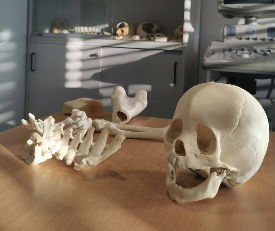
Natural haptic properties for surgical precision
Our models, 3D printed using a special process, are made of a gypsum composite material that is adjusted so that both handling and processing simulate real bone. This makes it possible to plan the operation realistically, and all steps of osteotomy, reduction and even screw placement can be precisely traced and tried out. This allows haptic training, efficient simulation and the use of the original instruments and screws in exactly the same way as they are used on the patient.
Thanks to its realistic weight and appearance, the model is also ideal for an informed discussion with the patient or family member in order to explain the necessary steps and to be able to show the result in advance.
Innovative solutions for special cases
We have developed many special solutions together with our customers. Your turn to challenge us!
Together with Prof. Dr. Roldán, we have developed a template plan for Treacher-Collins syndrome to reconstruct the zygomatic arch in just one step.
In order to make the lower jaw removable, but still fix it in its radiological position, we place magnets already during electronic model creation.
For a training on the special rotational fixator of the company Orthofix at the BGU Murnau, we realized bones with an internal cavity.
To depict tooth germs, we digitally realize them as a separate file and have them printed as red inclusions in a transparent model.
From virtual reconstruction to differential infiltration hardness gradients, we will make more out of your ideas.

